COMPREHENSIVE EYE CARE
A thorough eye exam is important for ensuring your long-term visual health.
‘Io Eye Center is committed to provide the highest standard of care for your eyes health. We treat a wide variety of ocular diseases and conditions. Please explore our options below and contact us for further evaluation and treatment consultation.
A comprehensive eye examination includes an evaluation for refractive changes affecting your eyesight through a process called refraction. This process fine-tunes the prescription of your glasses or contact lenses to give you the sharpest and clearest possible vision.
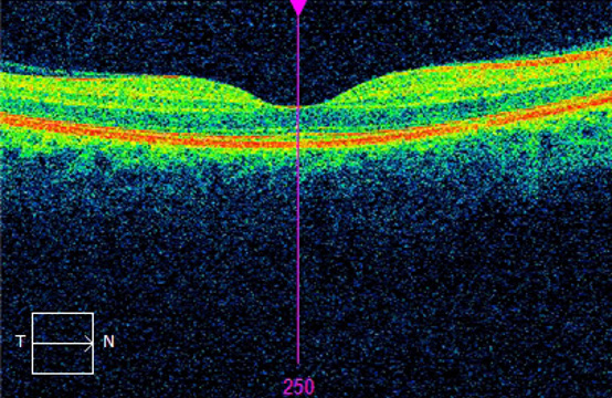
Your eye care physician evaluates the health of your eyes through the slit lamp, followed by a dilated fundus examination. Your eye doctor can actually see your eye's retinal blood vessels live in action. Certain conditions like high blood pressure or diabetes can affect the eye's blood vessels and your overall vision so eye exams are especially important for patients with these conditions. A dilated exam opens your pupil and allows you doctor to fully evaluate your lens, vitreous, optic nerve, blood vessels and permits a full view of your retina. This allows your doctor to diagnose eye diseases or risk factors for eye diseases like glaucoma or macular degeneration.
Click here for further information
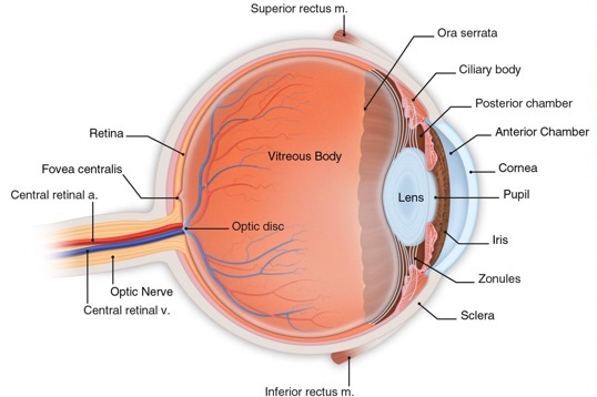
The parts of an eye are designed to fulfill their specific roles in the process of turning light into sight.
Cornea – The front “window” and main focusing element of the eye. It is made of a clear tissue that allows light to pass through without distortion.
Sclera – The sturdy white tissue that forms the outer wall of most of the eye.
Choroid – A dark tissue under the white sclera that helps to trap light within the eye. Blood vessels within it serve to nourish much of the eye.
Iris – The visible colored part of the eye that expands and contracts to let in light through its central opening for optimum vision.
Lens – The fine focusing element of the eye. It can vary its curvature to focus at different distances but starts to lose that full ability for most people between the age of 40 to 50.
Zonules – Flexible strands of tissue attached to the lens and the muscle of the ciliary body to facilitate changes in lens curvature.
Ciliary Body – A rim of tissue inside the eye that consists of a muscle to change the curvature of the lens and a gland-like element that produces aqueous humor.
Aqueous Humor – A clear solution produced in the ciliary body to provide nutrients for the front tissues of the eye and to maintain internal eye pressure. It circulates through the front chambers of the eye and drains into the bloodstream.
Retina – A photosensitive membrane that lines the back inside wall of the eye. It captures the images of light, changes them into electrical impulses, and sends the messages to the processing center of the brain for interpretation into sight.
Macula – The central, indented part of the retina where light rays, ideally, come to a focal point. It is 100 times more sensitive that the other parts of the retina to allow clear vision of detailed objects.
Optic Nerve – The nerve that carries electrical impulses from the eye to the processing center of the brain.
Optic Disc – The place on the retina where the optic nerve joins the back of the eye. This area is not sensitive to light.
Vitreous Gel – A clear, gel-like substance that occupies the main cavity of the eye.